THE EFFECTS OF THUMBSUCKING HABIT ON EXTERNAL NASAL MORPHOLOGY IN YOUNG MEN: A PHOTOGRAMMETRIC STUDY
Summary
Objective: In this study, we planned to determine primarily its effect on the craniofacial development and to measure the distance between the points, which were determined with the photogrammetric measurements over the nasal structures of the participants, who had thumb sucking habit up to 4 years of age in their childhood depending on the position of the hand and the pressure on the external nasal structures.Material and Methods: 20 male participants who have sucked their finger until at least 4 years of age and 30 healthy male volunteers who didn't have any thumb sucking habit were included in our study.
To ensure standardization, each person was photographed at the same distance (1 meter) from the same camera. Photogrammetric evaluation was performed blindly.
We took photos of each participant with the same camera at the same distance (1 m). Measurements were made from the front, lateral and basal profiles and the results were analysed.
Results: As a consequence of this study, nasal type width values were found to be significantly higher and the nose length was significantly shorter compared to the normal values.
Conclusion: These results indicated that ın nasal development period, morphology might be affected depending on many factors in people with thumb sucking behaviour.
Introduction
The thumbsucking behavior is one the most common and harmful habits. This habit is considered normal until 3-4 years of age in children. The habit that continues after this age is considered pathological and often occurs during emotional deprivation and nervous tension. The thumb is the most commonly sucked finger. The sucking of other fingers is relatively rare. If the thumb sucking behaviour cannot be prevented in a timely manner, it may cause an increase in overjet, anterior open-bite, narrow upper jaw and cross-bite. Deformations may also occur in the sucked fingers[1-3].Studies have demonstrated that most children break the thumb sucking habit before they were 5 years old. It was determined that both genders were equally affected and the degree of deformity was related to frequency, severity, duration of the sucking behaviour and on the intraoral position of the finger in the mouth[2,3]. Changes in the external nasal morphology occur due to the effects of factors such as high-arched palate, septum deviations or finger pressure in people who had exhibited the thumb sucking behaviour in their childhood.
The nose, which is at the center of the face, is one of the most important structure that affect the facial beauty. Specific and characteristic nasal forms are observed according to different race and geographical regions. Nasal values with different sizes, shapes and proportions were found in nasal anthropometric studies performed on different ethnic communities [5].
Anthropometric measurements are made between the points determined on the soft tissue or bone. Although there are no standardized nasal measurements in the morphological studies in the literature, there are different field measurements including nasal subunits, alar region and nasal type region[6,7]. The conventional anthropometric measurements are simple, inexpensive and they do not require complex equipment[8].
In this study, we aimed to compare the anthropometric values of the external nasal formations in persons, who exhibited thumb sucking behaviour in their childhood, with the volunteers who did not exhibit thumb sucking habit in their childhood or did not use a pacifier by using the computer aided photogrammetric measurements.
Methods
This study was approved by Medical and Health Sciences Research Committee of the Başkent University (KA18/57). As the participation in the study was based on a voluntary basis, an informed consent form was signed by all the volunteers participating in the study. 50 healthy young male aged between 18 and 25 were included in the study.
The participants were divided into two groups:
Group 1: Subjects, who exhibited thumb sucking behaviour up to 4 years of age. Group 2: Volunteers, who did not have thumb sucking or pacifier habit and had the below-mentioned criteria.
The criteria for inclusion in the study are as follows:
Being in the age range of 18-25, thumb sucking behaviour up to 3 years of age (for Group 1), no eye glasses in the childhood period (for Group 1), no previous intranasal/extranasal intervention or operation, no known cartilage bone related disease, having no nasal or facial trauma, does not have a syndrome that may cause craniofacial anatomic disorder, no diagnosis and treatment of cleft palate.
The exclusion criteria: Subjects who did not comply with the above-mentioned criteria were not included in the study.
Volunteers were selected among university students in Ankara. The students were asked about their history of finger-sucking in childhood. Those who had a history of finger sucking were asked to fill in demographic information.
Measurement:
To ensure standardization, each patient was photographed at the same distance (1.0 m from the camera) from the same standard digital camera (EOS 700D 18-55 DC III, Canon, Tokyo, Japan).
The photos were analyzed with the Adobe Photoshop (CC 2017, U 18.0.0, U.S.) and point-to-point and angular measurements were performed for the nasal analysis according to the literature. The photos were taken in front, both side and basal (close-up) views. When in the basilar view, the nasal type was connected with both side of the cornea9. All measurements were obtained by the same investigator (O.E.)
Points taken into consideration during the measurements:
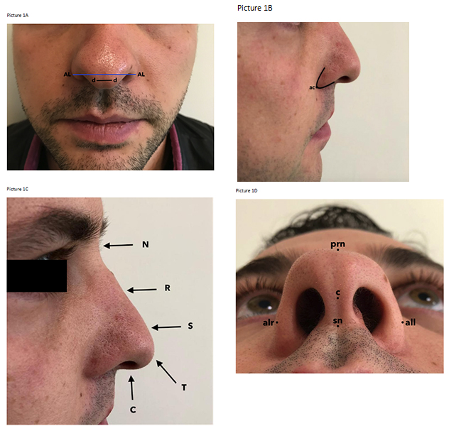
The soft landmarks were the pronasale (prn), the most anterior midpoint of the nasal tip; the top point of the columella (c); the subnasale (sn), the midpoint of the columella base at the columella-labial junction; and the alare (alr, all), the most lateral point of each alar contour9.
Seven standard anthropometric measurements were taken of the nasal region. Photogrammetric measurement distances: Front view; nose width (alr-all), dome width (d-d) (Picture 1A), Side view; Nasolabial angle (NL angle), nose length (N-T), nasal type protrusion (T-AL) (Picture 1B), Basal view; nasal base height (sn-prn), coumella length (c-sn) (Picture 1C).
The anatomic mark points: N: Nasion: The hollow point at the junction of the nose and the forehead, AL: The most curved point of each ala nosi. T: Tip: The most anterior point of the nasal tip, SN: Subnasale: The midpoint of the nasal base, d: Dome, PRN: Pronasale: The most anterior point of the nasal tip, C: The top point of the columella.
 Büyütmek İçin Tıklayın |
Picture 1: The anatomic mark points of the external regions of the nose. The front (1A), side (1B, 1C), basal (1D) views. N: Nasion, R: Rinion, S: Supratip, T: Tip, C: columella, A: Ala |
Statistical analysis
The data obtained from the subjects were uploaded to the software IBM SPSS Statistics v22 and analyzed. When evaluating the data, frequency distributions for categorical variables, descriptive statistics for numerical values (mean ± SD) are given. In order to choose the appropriate analysis method, primarily the normality test was applied to the numerical variables. If the normality assumption was provided, parametric tests were used; if not nonparametric tests were chosen. The average values of numeric data were calculated with the Student's t-test and the variables with non-normal distribution were evaluated with the Mann-Whitney U test. Regarding the comparative assessments, the accepted limit of significance was p<0.05.
Results
The study groups consisted of 20 male volunteers with thumb sucking history (Group 1) and 30 male volunteers without thumb sucking history (Group 2). Age distribution of the subjects is shown in Table 1.Table 1: Average age between groups 1 and 2.
The photogrammetric evaluation showed that the width of the nasal tip was significantly wider (p=0.025) and the nose length was significantly shorter (p=0.007) compared to the normal values.
Discussion
In the literature, it was stated that morphological changes in the teeth and the jaw structure might emerge in people with thumb sucking habit. The evaluation was based on the assumption that the possible skeletal anomalies and/or the contact and the pressure of other fingers on the external nasal tissues might occur during the thumb sucking. There are no similar studies in the literature. According to the results of our study, the width of the dome was significantly longer (p=0.025) and the nose length was significantly shorter (p=0.007) compared to the normal values.The skeletal development of the face, soft tissues and the muscular tissues take an important place in the face morphology. The nose sits on the midline of the face and plays an important role in the appearance of the face along with the chin and lips. It is also important in terms of the facial aesthetics and the respiratory function[10,11]. The development of the nose is completed in girls at the age of 16 and in boys at the age of 18. The studies focused on the nasal development showed that the nose grows about 1.5 mm per year in the forward and downward direction[11,12]. Regarding the results of our study, we concluded that the relatively shorter nose length in the thumb sucking people might depend on the restriction of the forward and downward development of the nose due to the pressure.
The shape and profile of the nose depends on both the bone and the cartilage components as well as the muscles on top and the integrity of the nose. The experimental studies on animals showed that the cartilage nasal septum has been shown to play an important role in the development of not only the nose but also the maxilla in animal experiments. Scott suggested that the cartilage is a primary growth center of the nasal septum and forms a force that pushes the front face down and forward[12-14]. Nasal morphology varies according to different ethnic groups and races[9]. Different nasal external analysis results were obtained in white and non-white race. These results provoked also the research on the variations between the nasal and other craniofacial structures[15,16].
The thumb sucking habit that continues after the early childhood may lead to certain physical and social problems. The anterior open bite is one of the most common problems in dentistry and the thumbs sucking habit is one of the etiological factors. Persistent thumb sucking and use of a pacifier prevent the development of an appropriate dental and alveolar process particularly in the anterior part. The morphological features of "long-face" are closely related to the anterior open bite. Furthermore, rotations in the opposite direction in the mandible and inadequate lip development are also observed[17].
In conclusion, our results (wider width of the dome and shorter nose length) showed that the nasal morphology might be affected in people with thumb sucking habit during the development period. Along with the anatomic problems in the tooth and jaw structure, the presence of high-arched palate and/or external finger pressure are factors that may be involved in the etiology. In addition, minor nose traumas during the childhood, allergic rhinitis or frequent infections may also play a role in these deformities. The small subject size is a limiting factor of our study. We believe that studies with larger and more homogeneous subject groups may confirm our results.
Acknowledgements
We would like to thank Tolgahan Yılmaz, Hilal Taylan Yılmaz, Rifat Ege Temel, Ahmet Turan, Zeynep Naz Uzun, Ozan Yaşar, the students of Başkent University Medical Faculty, who made it easier for us to reach University students and contribute to our study.
Reference
1) Anke B. The etiology of prolonged thumb sucking. Scand J Dent Res. 1971;79:54-59. [ Özet ]
2) Duncan K, McNamara C, Ireland AJ, Sandy JR. Sucking habits in childhood and the effects on the primary dentition: findings of the Avon Longitudinal Study of Pregnancy and Childhood. Int J Paediatr Dent. 2008;18:178-188. [ Özet ]
3) Figueiredo GC, Queiroga EC. Dystrophic calcinosis in a child with a thumb sucking habit: case report. Rev Hosp Clín Fac Med S Paulo 2000;55:177-180. [ Özet ]
4) Hein H. Thumb-suking as a cause of nasal septum deviation. Fortschr med. 1972:90(16):631-4. [ Özet ]
5) Zhenyu Y, Xiaoyan T and Jun F. Assessment of nasal base morphology using new proportion indices in Chinese. SpringerPlus. 2016;5:1275-86. [ Özet ]
6) Ofodile FA, Bokhari F, Ellis C. The black American nose. Ann Plast Surg 1993;31(3):209-218. [ Özet ]
7) Ofodile FA, Bokhari F. The African-American nose: part II. Ann Plast Surg 1995;34(2):123-1. [ Özet ]
8) Farkas LG, Philips JH, Katic M. Anthropometric anatomical and morphological nose widths in Canadian Caucasian adults. Can J Plast Surg. 1998;6(3):149-151.
9) He ZJ, Jian XC, Wu XS, Gao X, Zhou SH, Zhong XH. Anthropometric measurement and analysis of the external nasal soft tissue in 119 young Han Chinese adults. Craniofac Surg 2009;20(5):1347-51. [ Özet ]
10) Nehra K, Sharma V. Nasal morphology as an indicator of vertical maxillary skeletal pattern. J Orthod. 2009;36:160-6. [ Özet ]
11) Ferrario VF, Sforza C, Poggio CE, Schmitz JH. Three-dimensional study of growth and development of the nose. Cleft Palate Cranio fac J. 1997;34:309-17. [ Özet ]
12) Wisth PJ. Nose morphology in individuals with Angle Class I, Class II, or Class III occlusions. Acta Odontol Scand. 1975;33:53-7. [ Özet ]
13) Gungor AY, Turkkahraman H. Effects of airway problems on maxillary growth: A review. Eur J Dent.2009;3:250-4. [ Özet ]
14) Babula WJ, Jr, Smiley GR, Dixon AD. The role of the cartilaginous nasal septum in midfacial growth. Am J Orthod. 1970;58:250-63. [ Özet ]
15) Robison JM, Rinchuse DJ, Zullo TG. Relationship of skeletal pattern and nasal form. Am J Orthod 1986;89:499-506. [ Özet ]
16) Meng HP, Goorhuis J, Kapila S, Nanda RS. Growth changes in nasal profile from 7 to 18 years of age. Am J Orthod Dentofacial Orthop 1988;94:317-26. [ Özet ]
17) Oliveira AC, Pordeusb IA, Torresc CS, Martinsc MT, Paivab SM. Feeding and nonnutritive sucking habits and prevalence of open bite and crossbite in children/adolescents with Down syndrome. Angle Orthod. 2010;80(4):748-53. [ Özet ]


