UNILATERAL SECONDARY MIDDLE TURBINATE WITH SINUSITIS
2Fatih Üniversitesi Tıp Fakültesi, Radyoloji AD, Ankara, Türkiye
Introduction
Endoscopic nasal examination and radiological imaging techniques reveal anomalies of the nasal cavity such as septal deviation, concha bullosa, paradoxical middle turbinate and Haller cell. There are also other rare anomalies like secondary middle turbinate (SMT). It is known that SMT occurs bilaterally. Ostiomeatal unit obstruction and sinusitis due to SMT are very rare[1-4]. This paper describes a case of unilateral SMT which narrows the ostiomeatal unit and causes ethmoidal sinusitis.Case Presentation
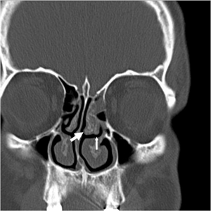
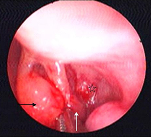
A 33 years old male presented with complaints of nasal obstruction, headache and snoring. Anterior rhinoscopy revealed nasal septum deviation and left inferior turbinate hypertrophy. Coronal computed tomography (CT) of the nasal cavity and paranasal sinuses demonstrated unilateral SMT (left) with anterior ethmoidal sinusitis (Fig. 1). Nasal endoscopy was performed; at the left side a structure which looks like a turbinate between the middle turbinate and lateral nasal wall was observed (Fig. 2).
 Büyütmek İçin Tıklayın |
Figure 1: Coronal computed tomography image, thick arrow indicates middle turbinate; and thin arrow, secondary middle turbinate |
 Büyütmek İçin Tıklayın |
Figure 2: Endoscopic view, black arrow indicates middle turbinate; white arrow, secondary middle turbinate; and star, uncinate process. |
Septoplasty and radiofrequency ablation of the inferior turbinates and endoscopic sinus surgery to the left side were performed under general anesthesia. SMT was removed and seen that it was attached to the bulla ethmoidalis. Bulla ethmoidalis was opened and anterior ethmoidectomy was performed. Anterior ethmoidal cells were filled with purulent discharge and the mucosa was hypertrophied. The patient had no complaint at the postoperative period.
Discussion
The SMT is a rare anomaly, first described by Khanobthamchai et al. as a bone, covered by soft tissue, originates from the lateral wall of the middle meatus[1]. We think that SMT is not a simple bony variation, it is an additional turbinate, as Aksungur et al. described[3].The SMT should be distinguished from the medial deviation and anterior folding of the uncinate process so called accessory middle turbinate[1]. In this case SMT and uncinate process were seen separately by endoscopy and CT.
The SMT was reported to be bilateral in all cases[1-4]. In our case SMT was detected unilaterally. For diagnosis of SMT, CT should be combined with nasal endoscopy otherwise, the diagnosis might be confused by other pathologic conditions. Some of the works in the literature depend only on CT findings and they might have been confused unilateral SMT with other pathologic conditions[2,3]. Consequently the incidence of unilateral SMT might be underestimated.
In the cases reported by Khanobthamchai et al. and Aykut et al. there were no ostiomeatal unit obstruction and sinusitis[1,2]. In the case of Apaydin et al. paranasal sinus mucosa was hypertrophied due to inferiorly and medially curving SMT, which were slightly narrowing the inferiorly lying hiatus semilunaris. In this case, SMT with its hypertrophic mucosa was narrowing hiatus semilunaris and at the same side there was ethmoidal sinusitis due to obstruction of the middle meatus.
As a result, other authors usually concluded that SMT is a bilaterally seen anatomical variation and is not a predisposing factor for sinusitis. However in this case we saw that SMT can occur unilaterally, can narrow ostiomeatal unit and can be a reason for sinusitis by narrowing ostiomeatal unit.
Reference
1) Khanobthamchai K, Shankar L, Hawke M, Bingham B: The secondary middle turbinate. J Otolaryngol. 1991;20:412-413 [ Özet ].
2) Aykut M, Gumusburun E, Muderris S, Adiguzel E: The secondary nasal middle concha. Surg Radiol Anat. 1994;16:307-309 [ Özet ].
3) Aksungur EH, Bicakci K, Inal M, Akgul E, Binokay F, Aydogan B, et al.: CT demonstration of accessory nasal turbinates: secondary middle turbinate and bifid inferior turbinate. Eur J Radiol. 1999;31:174-176 [ Özet ].
4) Apaydin FD, Duce MN, Yildiz A, Egilmez H, Ozer C, Talas UD: Inferomedially projecting pneumatised secondary middle turbinate. Eur J Radiol. 2002;43:42-44 [ Özet ].


