


HISTOPATHOLOGICAL EFFECTS OF LOCAL AND SYSTEMIC OZONE TREATMENT IN EXPERIMENTAL TYMPANIC MEMBRANE PERFORATIONS
2Bağcılar Eğitim Araştırma Hastanesi, Patoloji, İstanbul, Turkey
Summary
Aims: To investigate histopathological effects of local and systemic ozone application in experimental tympanic membrane perforations.Methods: Traumatic perforation was created by using a 21 gauge syringe needle under the microscope and then animals were then divided into 4 equal groups. The first group was the control group and it healed spontaneously. 0.5 mL local ozone oil was dropped to external ear in second group; 1.0 mL (1 µg / mL) ozone gas was given by rectal way in third group; 0.5 mL local ozone oil and 1.0 mL (1 µg / mL) rectal ozone gas was given in fourth group for 10 days. On day 11th, the tympanic membranes of the rats were excised and examined for inflammation, fibroblast proliferation, vascular density, collagenization and epithelization.
Results: It was observed that fibroblast proliferation, collagenization and epithelization scores of local, local+rectal, rectal ozone treatment groups were more significant compared to the control group (p<0.05).
Conclusion: This study showed the contribitions of local and systemic ozone treatment to wound healing by increasing inflammation, fibroblast proliferation, collagenization, vascular density and epithelization in experimental tympanic membrane perforations of rats.
Introduction
Tympanic membrane (TM) perforation is clinically common condition of people but the real incidence is unknown. The estimated incidence is reported below 1% all around the world, maybe this rate is low for estimates. The causes of perforation are primarily infections, followed by trauma. Traumas can be classified as penetrant trauma, barotrauma, acoustic trauma and iatrogenic trauma (myringotomy, ventilation tube insertion, ear canal clearance, ear lavage and hyperbaric oxygen therapy) [1].Small traumatic tympanic membrane perforations usually heal spontaneously. In some of these cases, the perforation doesn't close or partially closes with a thin membrane. However, many factors can lead to chronic perforation. In this situations, regeneration of the membrane is incompleted. Histologically, the squamous epithelium migrates through the perforation margin to the inner layer of the membrane. Theoretically, epithelialization of the wound edge stops spontaneous healing. Granulocyte Colony Stimulating Factor (G-CSF), Hepatocyte Growth Factor (HGF), Epidermal Growth Factor (EGF), Gelfoam, Saline irrigation, Ascorbic acid (C vit), Hyaluronic acid were used to accelerate TM repair in experimental studies [2-5].
Previous studies showed ozone treatment accelerates wound healing by increasing growth factors such as VEGF, TGF-B and PDGF. In this study, we thought ozone therapy would increase the wound healing at a cellular level in traumatic tympanic membrane perforations.
Methods
This study started after confirmation of Experimental Animal Ethics Board dated on 26.01.2016 and numbered 2016/04. 20 healthy Wistar Hannover genus and male rats about 335 Gr (250-450) weight were used in the study. The rats lived at 16-21C temperature, 50% moisture and fed with Pellet. Our study complied on the part of animal care of Helsinki declaration.Ketamine hydrochloride (Ketalar; Pfizer Drug, Istanbul, Turkey) 60 mg / kg intramusculer (i.m) and xylazine hydrochloride (Rompun; Bayer Healthcare, Kansas, USA) 12 mg / kg / intramusculer (i.m) were used for anesthesia of rats prior to myringotomy. Then, we examined the 40 ears of 20 rats on microscope. We removed debris and cerumen from the external auditory canal. Healty tympanic membrane images were detected in all rats . Traumatic perforation approximately 1 mm wide was created by using a 21 gauge syringe needle through anterior quadrant of both tympanic membranes of the rats. Then, animals were divided into 4 equal groups. The first group was the control group and it healed spontaneously. 0.5 mL local ozone oil (Arozon ozonated olive oil; Ar-Tekin Drug, Çankırı, Turkey) was dropped to external auditory canal in second group; 1.0 mL (1 µg / mL) ozone gas was given by rectal way in third group; 0.5 mL local ozone and 1.0 mL (1 µg / mL) rectal ozone gas were given in fourth group for 10 days. On first day of treatment, one of the rats in local ozone group; on second day of treatment, one rat in rectal group and one rat in rectal+local ozone groups were died and seventeen rats were treated completely. On the 11th day of the study, the animals were sacrified after injection of 80 mg / kg / intramusculer (i.m) ketamine hydrochloride and 20 mg / k.g / i.m. xylazine hydrochloride. Subsequently, temporal bone dissection of the rats was carried out for histopathological examination.
2 of the 10 temporal bones in the 5 control group rats, 2 of the 8 temporal bones in 4 local ozone group rats, 1 of the 8 temporal bones in 4 rectal ozone group rats and 1 of the 8 temporal bones in the 4 rectal + ozone group rats were excluded from the study since tympanic membranes were damaged during dissection, 28 tympanic membranes with tympanic bones could be excised. Then, the tissues were placed in a 10% formaldehyde solution and sent to the pathology. Histopathological examination of tissue specimens were performed without knowledge the test groups. Vascularization (vessel density), fibroblast proliferation, collagenization and epithelialization scores were evaluated as 0: none 1: mild 2: middle 3: severe. Inflammation severity scoring system of Hooker et al was used as 0: no inflammation, 1: rare lymphocytes and plasma cells, 2: increased lymphocyte, neutrophil and plasma cells, 3: a large number of mixed inflammatory cells and micro abscesses [6].
Statistical analyzes were performed using the NCSS (Number Cruncher Statistical System) 2007 Statistical Software (Utah, USA) package program . Descriptive statistical methods (mean, standard deviation, median and interquartile range), Kruskal Wallis test in group comparisons, Dunn's multiple comparison test in subgroup comparisons were used to evaluate the data. The results were evaluated at a significance level of p <0.05.
Results
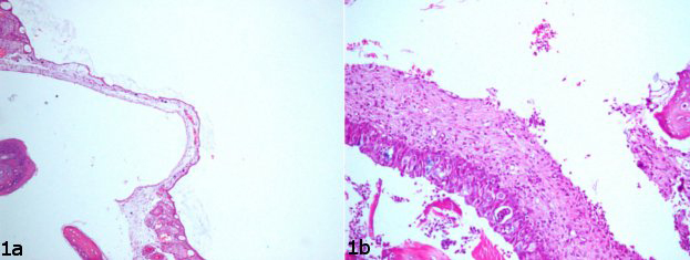
İnflamation severityThe mean inflammation scores of control, local, rectal and local+rectal treatment groups were orderly 1.38±0.52, 2±0, 1±0, 2.14±0.38 (Table 1). Inflammation values of local + rectal ozone therapy group were found to be significantly higher than the control and rectal ozone treatment groups (p = 0,011, p = 0,0001). The inflammation values of local ozone treatment group were found to be significantly higher than the control and rectal ozone treatment groups (p = 0,02, p = 0,0001). No statistically significant difference was observed between the other groups (p> 0,05) (Table 2). Mild inflammation was observed in control group and severe inflammation was observed in the local + rectal ozone therapy group (Figure 1. a,b).
)
)
 Büyütmek İçin Tıklayın |
Figure 1: a,b. Mild inflammation (a) was observed in control group and severe inflammation (b) was observed in the local + rectal ozone therapy group. |
Vascular density
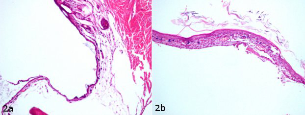
The mean vascular density scores of control, local, rectal and local+rectal treatment groups were orderly 1.25±0.46, 2.17±0.75, 2±0 and 3±0 (Table 1). Vascular density values of the local + rectal ozone treatment group were found to be statistically significantly higher than the control , local ozone treatment and rectal ozone treatment groups (p = 0,0001, p = 0,014, p = 0,0001). No statistically significant difference was observed between the other groups (p> 0,05) (Table 2). Random vascular structures were observed in control group and severe vascularization was observed in local+rectal ozone treatment group (Figure 2. a,b).
 Büyütmek İçin Tıklayın |
Figure 2: a,b. Random vascular structures (a) were observed in control group and severe vascularization (b) was observed in local+rectal ozone treatment group. |
Collagenization
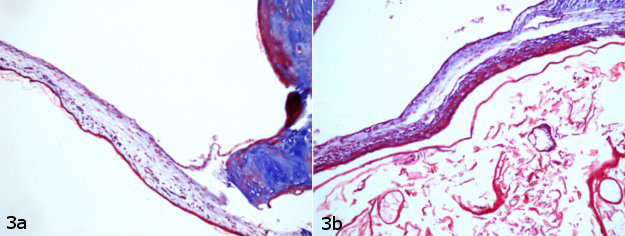
The mean collagenization scores of control, local, rectal and local+rectal treatment groups were orderly 1.50±0.54, 2.17±0.41, 2.29±0.49 and 2.57±0.54 (Table 1). The collagenization values of the control group were statistically significantly lower than the local ozone treatment, rectal ozone treatment and local + rectal ozone treatment groups (p = 0,032, p = 0,017, p = 0,006). No statistically significant difference was observed between the other groups (p> 0,05) (Table 2). Collagen density showed mild staining in lamina propria in the control group. In contrast, local+rectal ozone treatment group had severe collagenization in lamina propria (Figure 3. a,b).
 Büyütmek İçin Tıklayın |
Figure 3: a,b. Control group had mild collagenization (a) in lamina propria and local+rectal ozone treatment group had severe collagenization (b) in lamina propria. |
Fibroblast proliferation
The mean fibroblast proliferation scores of control, local, rectal and local+rectal treatment groups were orderly 1.38±0.52, 2.17±0.41, 2.29±0.49 and 2.57±0.54 (Table 1). The fibroblast proliferation values of the control group were statistically significantly lower than those of local ozone treatment, rectal ozone treatment and local + rectal ozone treatment groups (p = 0,015, p = 0,008, p = 0,004). No statistically significant difference was observed between the other groups (p> 0,05) (Table 2). Control group had a little fibroblast in lamina propria and many fibroblasts were observed in local+rectal ozone treatment group (Figure 4. a,b).
 Büyütmek İçin Tıklayın |
Figure 4: a,b. Control group had a little fibroblast (a) in lamina propria and many fibroblasts (b) were observed in local+rectal ozone treatment group. |
Epithelization
The mean epithelization scores of control, local, rectal and local+rectal treatment groups were orderly 1.75±0.89, 2.17±0.41, 2.29±0.49 and 2.57±0.54 (Table 1). The epithelialization values of the control group were statistically significantly lower than local ozone treatment, rectal ozone treatment and local + rectal ozone treatment groups. (p = 0.042, p = 0.037, p = 0.01). No statistically significant difference was observed between the other groups (p> 0,05) (Table 2). Epithelization was observed as a thin layer in control group and full layer epithelization was observed in local+rectal ozone treatment group (
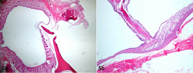
Büyütmek İçin TıklayınFigure 5: a,b. Epithelization was observed as a thin layer (a) in control group and full layer epithelization (b) was observed in local+rectal ozone treatment group.
Discussion
Wound healing of the tympanic membrane is common in the initial hemostatic and inflammatory stages due to vascular distribution; but it is completely different from other tissues at the proliferation and migration phases. In many wound healing, granulation tissue is formed as a platforma deposit that helps epithelization progress. In typmanic membrane regeneration, rebuilding of the fibrous component follows forming bridges of squamous epithelium layer over wound. Thickened lamina propria contains type 1 and type 3 collagen [7]. The original tympanic membrane contains predominantly type 2 collagen [8].When tympanic membrane perforations heal spontaneously with neomembrane, the fibrous layer is deficient. Thus, suboptimal healing may lead to the formation of retraction pockets [9]. In the treatment of ozone, there was a strong middle layer associated with dense collagenization and fibroblast proliferation. Ozone treatment has similar effects with the research of Yeo et al. They also reported PDGF-AA treatment which increases fibroblast number in the middle layer [10]. So, ozone therapy provides original tympanic membran formation with 3 layer.
Ozone is a triatomic molecule consisting of 3 oxygen atoms. It has a unstable structure and may convert to molecular oxygen and oxygen atoms. Ozone does not affect by entering into cells. It forms free radicals such as hydrogen peroxide and polyunsaturated fatty acids (PUFAs) in cell membranes. Thus, it increases transcription factors like nuclear factor-kappa B (NF-kB) for synthesis of growth factors and sustaining cell viability [11].
Topical ozone application has three main forms; ozone water, ozone oil and ozone gas. Ozone treatment is a cost effective and it has less side effect. As it is used in a wide range of biological applications, it is known that activates the immune system to show antimicrobial effect and increase wound healing [12]. On the other hand, ozone treatment is not common in otorhinolaryngology practice. There are clinical trials with ozone therapy which show effectivity of ozone in sudden hearing loss [13,14]. In contrast, it is not effective in the treatment of tinnitus [15].
Ozone application is beneficial for wound healing. Kim et al. showed the acceleration of healing process in full thickness skin wounds due to administiration of ozone oil [16]. In study reported by Erginel et al. the accumulation of collagen in the anastomosis line was observed more in the ozone groups than in the control group after colonic anastomosis [17]. In our study, we showed likely that inflammation, fibroblast proliferation, vascular density, collagenization and epithelialization scores were the highest in the local + rectal ozone treatment groups.
Granulocyte Colony Stimulating Factor (G-CSF), Hepatocyte Growth Factor (HGF), Epidermal Growth Factor (EGF), Gelfoam, Saline irrigation, Ascorbic acid (C vit), Hyaluronic acid were used for experimental studies to accelerate TM repair. Aissa et al. showed gelfoam or G-CSF did not affect to tympanic membrane healing [9]. In the study reported by Güneri et al. , there was no statistically significant difference in tympanic membrane thickness, fibroblastic reactions and neovascularization between the 3 groups which are treated with hyaluronic acid, EGF and mitomycin in experimental tympanic membrane perforations [18]. In the experimental study reported by Erkilet et al. , histopathological evaluation of tympanic membrane healing of PRP group was earlier than the control group, but there was no statistically significant difference between two groups in terms of lamina propria edema, neovascularization fibroblastic reaction and inflammatory cell hyperplasia [19]. Similarly, Alan et al. found no significant difference inflammation, epithelial migration and neovascularization on tympanic membrane of ginea pigs between the intraperitoneal pentoxifylline group and intraperitoneal saline group [20]. In the study reported by Gunes et al, there was no statistically significant difference in terms of inflammation and lamina propria edema between control group and ascorbic acid treatment group [21]. In contrastly, we found statistically increase of vascularization, fibroblast proliferation, and epithelization due to experimental ozone therapy in this study.
Inflammation is the first phase of wound healing and occurs within the first 3-4 days. In our study, severe inflammation reached to the 10th day in ozone treatment groups. Propably, it adversely affect perforation closure due to block passing to another phase. Remodeling phase starts in 3 weeks so our study didn't include remodeling phase. There is a need for studies that include ozone treatment for 3 weeks and evaluates the perforation diameter by microscopic examination.
Conclusion
This study showed the contribitions of local and systemic ozone treatment to wound healing by increasing inflammation, fibroblast proliferation, collagenization, vascular density and epithelization in experimental tympanic membrane perforations of rats.
ACKNOWLEDGEMENTS
We have started to this project after the confirmation of Bagcilar Training and Research Hospital Experimental Animal Ethics Board dated 29.03.2016 and numbered 2016/04. There is no conflict of interest and financial disclosure.
Reference
1) Cohen D, Tamir D. The prevalence of middle ear pathologies in Jerusalem school children. -Am J Otol. 1989;10:456-9. [ Özet ]
2) Laurent C, Hellstrom S, Fellenius E. Hyaluronan improveions the healing of experimental tympanic membrane perforations. A comparison of preperations with different rheologic properties. Arch. Otolaryngol. Head Neck Surg. 1988;114:1435-41. [ Özet ]
3) Spandow O, Hellstrom S. Healing of tympanic membrane perforation-a complex process influenced by a variety of factors. Acta. Otolaryngol. Suppl. 1992;492:90-3. [ Özet ]
4) Amoils CP, Jackler RK, Lustig LR. Repair of chronic tympanic membrane perforations using epidermal growth factor. J. Otolaryngol. Head Neck Surg. 1992;107:669-83. [ Özet ]
5) Kaftan H, Reuther L, Miehe B, Hosemann W, Beule A. Inhibition of fibroblast growth factor receptor 1: influence on tympanic membrane wound healing in rats. Eur. Arch. Otorhinolaryngol. 2012;269:87-92. [ Özet ]
6) Hooker GD , Taylor BM, Driman DK. Prevention of adhesion formation with use of sodium hyaluronate-based bioresorbablemembrane in a rat model of ventral hernia repair with polypropylene mesh--a randomized, controlled study. Surgery. 1999;125:211-6. [ Özet ]
7) K. Stenfeldt, C. Johansson, S. Hellstro¨m. The collagen structure of the tympanic membrane: collagen types I II, and III in the healthy tympanic membrane, during healing of a perforation, and during infection. Arch. Otolaryngol. Head Neck Surg. 2006;132:293-8. [ Özet ]
8) J. Knutsson, D. Bagger-Sjoback, M. Von Unge. Distribution of different collagen types in the rat's tympanic membrane and its suspending structures. Otol. Neurotol. 2007;28:486-91. [ Özet ]
9) Hong P, Bance M, Gratzer PF. Repair of tympanic membrane perforation using novel adjuvant therapies: A contemporary review of experimental and tissue engineering studies. Int. J. Pediatr. Otorhinolaryngol. 2013;77:3-12 [ Özet ]
10) Yeo SW, Kim SW, Suh BD, Cho SH. Effects of Platelet-Derived Growth Factor-AA on the Healing Process of Tympanic Membrane Perforation. Am. J. Otolaryngol. 2000;21:153-60. [ Özet ]
11) Valacchi G, Sticozzi C, Pecorelli A, Cervellati F, Cervellati C, Maioli E. Cutaneous responses to environmental stressors. Ann. N. Y. Acad. Sci. 2012;1271:75-81. [ Özet ]
12) Borges GÁ, Elias ST, Da Silva SMM, Magalhães PO, Macedo SB, Ribeiro APD et al. In vitro evaluation of wound healing and antimicrobial potential of ozone therapy. J. Craniomaxillofac. Surg. 2017;45:364-370. [ Özet ]
13) Ragab A, Shreef E, Behiry E, Zalat S, Noaman M. Randomised, double-blinded, placebo-controlled, clinical trial of ozone therapy as treatment of sudden sensorineural hearing loss . J. Laryngol. Otol. 2009;123:54-60. [ Özet ]
14) Tasdoven GE, Derin AT, Yaprak N, Ozcaglar HU. The place of hyperbaric oxygen therapy and ozone therapy in sudden hearing loss. Braz. J. Otorhinolaryngol. 2017;83:457-63. [ Özet ]
15) Sonmez O, Kulahlı I, Vural A, Sahin MI, Aydın M. The evaluation of ozone and betahistine in the treatment of tinnitus. Eur. Arch. Otorhinolaryngol. 2013;270:1999-2006. [ Özet ]
16) Kim HS, Noh SU, Han YW, Kim KM, Kang H, Kim HO et al. Therapeutic Effects of Topical Application of Ozone on Acute Cutaneous Wound Healing. J. Korean Med. Sci. 2009;24:368-74. [ Özet ]
17) Erginel B, Erginel T, Aksoy B, Dokucu Aİ. Effect of Ozone Therapy (OT) on Healing of Colonic Anastomosis in a Rat Model of Peritonitis. Balkan Med J. 2014;31:249-53. [ Özet ]
18) Guneri EA, Tekin S, Yilmaz O, Ozkara E, Erdag TK, İkiz AO. The Effects of Hyaluronic Acid, Epidermal Growth Factor, and Mitomycin in an Experimental Model of Acute Traumatic Tympanic Membrane Perforation. Otol. Neurotol. 2003;24:371-6. [ Özet ]
19) Erkilet E, Koyuncu M, Atmaca S, Yarim M. Platelet-rich plasma improves healing of tympanic membrane perforations: experimental study. J. Laryngol. Otol. 2009;123:482-7. [ Özet ]
20) Lim AA, Washington AP, Greinwald JH, Lassen LF, Holtel MR. Effect of pentoxıfylline on the healing of guinea pig tympanic membrane. Ann. Otol. Rhinol. Laryngol. 2000;109:262-6. [ Özet ]
21) Gunes A, Mutlu M, Akin I, Koybasioglu F, Guvey A, Karasu MF et al. The Impact of Systemic and Local Administration of Ascorbic Acid on Traumatic Perforation of Tympanic Membrane and Myringosclerosis. J. Int. Adv. Otol. 2015;11:48-52. [ Özet ]