


DOES THE HOUNSFIELD UNIT MEASURED IN MULTIPLE FOCUS IN HIGH RESOLUTION TEMPORAL BONE TOMOGRAPHY CONTRIBUTE TO THE DIAGNOSIS OF OTOSCLEROSIS?
2İstanbul Arel Üniversitesi Tıp Fakültesi, Kulak Burun Boğaz Anabilim Dalı İstanbul, Turkey
3Silopi Devlet Hastanesi Kulak Burun Boğaz, Şırnak, Turkey
4Dicle Üniversitesi Tıp Fakültesi Kulak Burun Boğaz Ana Bilim Dalı, Diyarbakır, Turkey
Summary
Purpose: In this study, it was aimed to investigate the function of high-resolution computed tomography (HRCT) densitometry in the diagnosis of otosclerosis by performing densitometric measurements on the foci around the cochlea.Methods: The HRCT images of 0.5 mm thin-slice axial sections were obtained from 30 patients with otosclerosis, who had been surgically confirmed and performed computed tomography of the temporal bone before the surgery, and 30 patients, who had undergone cochlear implant surgery and had been identified as the control group were examined. Densitometric measurements obtained from 7 different points of the otic capsule were recorded as the mean Hounsfield unit (HU) for each patient.
Results: When we compared the patient and the control groups, a statistically significant difference was found in densitometric measurements in 6 of the 7 different regions (p<0.05). There was no statistically significant difference only in the HU 4 region (p>0.05). There was also a statistically significant difference between these two groups in the mean HU value obtained by measurements performed in 7 regions (p<0.05). In addition, no significant difference was found between both ears in all regions in the densitometric measurements at 7 different points of the otic capsule between the operated and non-operated ears of these patients (p> 0.05).
Conclusion: In our study, it was found that the density around the otic capsule was lower in patients with otosclerosis compared to normal patients.
Introduction
Otosclerosis is osteodystrophy of the otic capsule with progressive hearing loss. Histologically, it is characterized by abnormal bone resorption and deposition in the otic capsule and is seen only in humans [1]. In cases of otosclerosis, the lesions can be spongiotic, fibrotic, and sclerotic, histologically[2]. Otosclerotic foci may develop from a very small focus in the anterior crus or completely obliterating the stapes' base. The "Fissula ante Fenestram", which is located in the immediate anterior of the oval window, is most frequently involved in the temporal bone[3]. Otosclerosis can be diagnosed based on detailed anamnesis, physical examination, and audiological findings. Computed tomography (CT) is generally preferred as the imaging method in otosclerosis. CT has an important role in the diagnostic evaluation of patients with otosclerosis and cochlear otosclerosis, and the selection of the ear to be operated[4]. High-resolution computed tomography (HRCT) has a significant role in imaging the labyrinthine and bony capsule of the temporal bone. The extent of otosclerosis into the cochlear capsule can be quantitatively evaluated using densitometric measurements[5]. With high-resolution computed tomography (HRCT), the spread of otosclerosis to the cochlear capsule can be evaluated by densitometric measurements of the otosclerotic foci. In this study, we aimed to investigate the function of high-resolution computed tomography (HRCT) densitometry in the diagnosis of otosclerosis by performing densitometric measurements on the foci around the cochlea in patients with otosclerosis, who had been surgically confirmed.Methods
PatientsIn this study, 30 patients with surgically confirmed otosclerosis (otosclerosis group) and 30 patients who underwent cochlear implant surgery (control group) were examined. Department of Otorhinolaryngology between January 2013 and December 2019. In the otosclerosis group, the evaluation was made on the operated ears with confirmed otosclerosis. Patients over 18 years of age, who presented to our clinic with severe post-lingual bilateral hearing loss and underwent cochlear implant surgery were evaluated as the control group. HRCT images obtained before the surgery were evaluated for each patient. In the otosclerosis group, patients with a history of surgery performed for non-auricular reasons were excluded from the study. Implant patients with completely normal inner and middle ear structures on HRCT were selected as the control group. Approval was received from the Ethics Committee of the Faculty of Medicine of ?. University (147, dated 25.02.2021).
Radiological Evaluation
The HRCT images of 0.5 mm thin-slice axial sections obtained from the 60 patients included in this study were examined. Densitometric measurements obtained from 7 different points of the otic capsule were indicated as the mean Hounsfield unit for each patient (Figure 1). The patients included in the study were evaluated with 64-slice high-resolution CT (Brilliance CT 64 Philips Medical Systems, Cleveland, OH) in our hospital. Temporal CT imaging was performed axially for both ears with a slice thickness of 0.67 mm, at a tube voltage of 120kV and effective mAs of 112 according to the standard protocol. The axial images were reconstructed in the coronal and sagittal planes by the technician and submitted to the workstation. The images obtained were evaluated using PACS (picture archiving and communication system) (Infinitt, Korea).
 Büyütmek İçin Tıklayın |
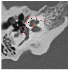
Figure 1: CT scan (axial view) of a left temporal bone in a patient with otosclerosis showing the different anatomical points at which the bone density was measured 1= anterior of the oval window (behind the intersection of the stapes posterior arch and the stapes footplate); 2= posterior of the oval window (in front of the intersection of the stapes anterior arch and the stapes footplate); 3= lateral to the mid-turn of the cochlea; 4= lateral to the mid-turn of the cochlea with the apex of the cochlea; 5= apex of the cochlea; 6= anteromedial of the middle turn of the cochlea; 7= anteromedial of the basal turn of the cochlea. |
Statistical Analysis
IBM SPSS 21.0 for Windows statistical package software was used for the statistical evaluation of the data obtained in our study. The measurement variables were presented as mean ± standard deviation (SD), and the categorical variables were presented as numbers and percentages (%). The data were evaluated for the normality of distribution. The independent t-test was used for the comparison between the two groups with normal distribution. Spearman's correlation analysis was used for analyzing the correlations between the variables that did not conform to a normal distribution. The qualitative variables were compared using the Chi-square (χ2) test. The comparison of the paired groups with non-normal distribution was performed using the Mann-Whitney U test. Hypotheses were considered two-sided, and p≤0.05 was accepted as a statistically significant result.
Results
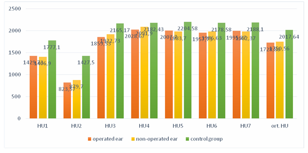
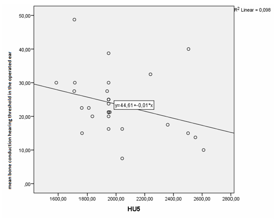
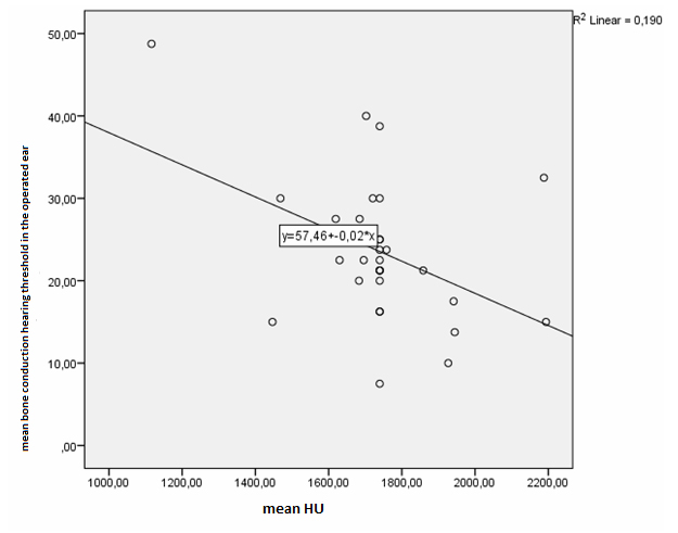
This study examined 30 patients with otosclerosis, who had been surgically confirmed, and 30 patients who underwent cochlear implant surgery (control group) were examined. Of the patients with otosclerosis, 15 were male (50%), and 15 were female (50%). In the otosclerosis group, the mean age was 37.17± 9.70 years. There were 16 female (53.3%) and 14 male (46.7%) patients in the control group. The mean age of the control group was 34.73 ± 13.84 years. There was no difference in the statistical distribution of the patient and control groups in terms of gender and age (p>0.05) (Table 1, Table 2). The mean air conduction thresholds of the patient group with otosclerosis were 57.94 ± 12.29 dB for the operated ear and 38.04 ± 14.13 dB for the non-operated ear. The bone air conduction thresholds of these patients were 23.54 ± 8.81 dB in the operated ear and 18.58 ± 7.99 dB in the non-operated ear (Table 3). The mean Hounsfield unit (HU) values obtained in densitometric measurements from 7 different regions of the cochlea for the axial section in the HRCT of the otosclerosis and control groups were presented in Table 4. When we compared the patient and the control groups, we found a statistically significant difference in densitometric measurements in 6 of the 7 different regions (p<0.05). We found no statistically significant difference only in the HU 4 region (p>0.05). We also found a statistically significant difference between these two groups in the mean HU value obtained in the measurements performed in 7 regions (p<0.05) (Table 4) (Chart 1). In addition, no significant difference was found between the two ears in all regions in the densitometric measurements we performed using HRCT at 7 different points of the otic capsule between the operated and non-operated ears of these patients (Table 5) (Chart 1). Moreover, a negative and significant relationship was found between the air conduction hearing threshold and the mean HU values in 7 regions according to the result of the Spearman correlation test we conducted to determine the relationship between the HU values in 7 regions and, the audiometric air conduction hearing thresholds and the audiometric bone conduction hearing thresholds of patients with otosclerosis. We found a negative (r=0.383, p=0.037) and significant relationship between the HU4, HU5, and mean HU values and the bone conduction hearing threshold. No significant relationship was found in the other regions (Table 6) (Chart 2, Chart 3, Chart 4).)
)
)
)
)
)
Table 1: Gender variable data of the groups
Table 2: Data regarding the age variable
Table 4: Comparison of the HU value belonging to the otosclerosis and control groups
Table 5: Comparison of the HU values belonging to the operated and non-operated ear groups.
 Büyütmek İçin Tıklayın |
Chart 1: Mean values of the HU parameters belonging to the operated and non-operated groups, and the control group. |
 Büyütmek İçin Tıklayın |
Chart 2: Correlation between HU4 and mean bone conduction hearing threshold in the operated ear |
 Büyütmek İçin Tıklayın |
Chart 3: Correlation between HU5 and mean bone conduction hearing threshold in the operated ear |
 Büyütmek İçin Tıklayın |
Chart 4: Correlation between mean HU value and mean bone conduction hearing threshold in the operated ear |
Discussion
Otosclerosis is a disease, which involves bone dystrophy. Histologically, bone is absorbed by osteoclastic activity and new bone is deposited by osteocytes. In the active phase, the lesions contain irregular bones that are rich in low mineralization osteocytes and highly vascularized connective tissue. There are no pathological findings except otosclerosis on CT in the majority of the patients with otosclerosis. The location and size of demineralized lesions vary. Some lesions appear as sclerotic lesions, which are less vascular and more solid. Of sclerotic lesions, 70-90% are located near the Fissula ante Fenestram (FAF)[6]. In their histological study, Schuknecht and Barber detected otosclerotic foci at the FAF in 123 temporal bones in 118 patients[7].It is difficult to find sclerotic lesions due to their small size and density close to normal bone. Therefore, the density measurements by CT appear valuable as they provide the potential to quantify the presence of otosclerosis using CT. Grayeli et al. conducted a study on 10 patients with otosclerosis and 33 control patients with vestibular schwannoma and found that the bone density they measured at seven different points of the otic capsule on 0.5 mm thin-section CT was significantly low around the Fissula ante Fenestram[8]. Zhu M. et al. conducted a study on 34 patients with otosclerosis and 33 control patients and detected lower intensity in the areas anterior to the oval window (ROI 1) and posterior to the oval window (ROI 2) compared to the control group in the densitometric measurements by HRCT from 7 areas around the otic capsule[9]. Kawase et al. found that the density in the anterior of the Fissula ante Fenestram and the internal acoustic canal from 8 different points of the otic capsule was significantly lower in patients with otosclerosis compared to the control group[10]. In their study, Kutlar G. et al. detected statistically significantly lower density in otosclerotic ears compared to the control group only in the FAF area in the densitometric measurements performed at 8 different points of the otic capsule[11]. Puigross et al. stated that bone densities in Hounsfield units (HU) from 28 otosclerotic ears were compared to the densities of 33 non-otosclerotic capsules. These densities were measured in eight regions of interest (ROI) where the otosclerotic foci are usually found and the density in the fissula ante fenestram (ROI1) and in the precochlear region (ROI3) seem to be the most valuable parameters to make a diagnosis of otosclerosis.[12]
In our study, we performed densitometric measurements at seven different points of the otic capsule with thin-section CT between patients with otosclerosis, who had been surgically confirmed, and the patients, who underwent cochlear implant surgery (control group) were examined. We measured patients with otosclerosis significantly lower at six different points compared to the patients in the control group (p<0.05). When we compared with the literature, we found that the density in the overall otic capsule was lower in patients with otosclerosis, unlike other studies. In addition, we found that the mean density measurement we performed yielded significantly lower results compared to the patients in the control group. We believe that densitometric measurements by HRCT have a place in the diagnosis of otosclerosis. However, our results should be supported by studies to be conducted with larger numbers of patients. In otosclerosis, there is usually bilateral hearing loss, and one of the ears is asymmetrical as one ear begins before the other[13,14] The patients who were operated in our study had all been operated on one ear. Our selection criterion for the ear to be operated included the selection of the ear with more severe hearing loss in terms of hearing. In the densitometric measurements we performed with thin-section CT at 7 different points of the otic capsule on the operated and non-operated ears of these patients, there was no statistically significant difference between the two ears at all regions. Swartz et al. demonstrated a correlation between decalcification of the otic capsule and sensorineural hearing loss at high, moderate, and low frequencies. However, they did not make a quantitative evaluation such as densitometry[15] Guneri et al. was described a strong correlation between the site of the otosclerotic focus around the cochlea and the audiometric frequency of the sensorineural HL[16]. Kawase et al. found a negative correlation between densitometric values and bone conduction threshold in the ROI 2, ROI5, and ROI6 areas of the otic capsule[10] . Zhu M et al. stated that there was a positive correlation between the mean ROI1 value and the air-bone conduction in their study[9]. In our study, a statistically negative correlation was found between the bone conduction hearing threshold and the mean density value in the HU4, HU5 and mean HU areas in patients with otosclerosis. No statistical significance was determined in other areas. There was no statistically significant correlation between the air conduction hearing threshold and density value.
In general, we found that the density around the otic capsule was lower in patients with otosclerosis compared to normal patients. In accordance with the literature, we believe that densitometric measurements of thin-section CT are useful in the diagnosis of patients with otosclerosis.
Reference
1) HYPERLINK "https://pubmed.ncbi.nlm.nih.gov/?term=Makarem+AO&cauthor_id=20195188" Andre O Makarem HYPERLINK "https://pubmed.ncbi.nlm.nih.gov/20195188/" l "affiliation-1" o "Histopatoloji Anabilim Dalı, House Ear Institute, UCLA Medical Center, Los Angeles, California, ABD" 1 , HYPERLINK "https://pubmed.ncbi.nlm.nih.gov/?term=Hoang+TA&cauthor_id=20195188" Thu-Anh Hoang , HYPERLINK "https://pubmed.ncbi.nlm.nih.gov/?term=Lo+WW&cauthor_id=20195188" William WM Lo , HYPERLINK "https://pubmed.ncbi.nlm.nih.gov/?term=Linthicum+FH+Jr&cauthor_id=20195188" Fred H Linthicum Jr. , HYPERLINK "https://pubmed.ncbi.nlm.nih.gov/?term=Fayad+JN&cauthor_id=20195188" Jose N Fayad . Cavitating otosclerosis:clinical, radiologic, and histopathologic correlations. Otol Nörotol.2010 Nisan;31(3):381-4. [ Özet ]
2) HYPERLINK "https://pubmed.ncbi.nlm.nih.gov/?term=Linthicum+FH+Jr&cauthor_id=8341566" FH Linthicum Jr. Histopathology of otosclerosis. Otolaryngol Kliniği Kuzey Am .1993 Haziran;26(3):335-52. [ Özet ]
3) HYPERLINK "https://pubmed.ncbi.nlm.nih.gov/?term=Bretlau+P&cauthor_id=5360033" P Bretlau J. Relation of the otosclerotic focus to the fissula ante-fenestram. Laryngol Otol.1969 Dec;83(12):1185-93. [ Özet ]
4) HYPERLINK "https://pubmed.ncbi.nlm.nih.gov/?term=Naumann+IC&cauthor_id=16240935" Ilka C Naumann HYPERLINK "https://pubmed.ncbi.nlm.nih.gov/16240935/" l "affiliation-1" o "Department of Otolaryngology-Head and Neck Surgery, Indiana University, Indianapolis, Indiana, USA." 1 , HYPERLINK "https://pubmed.ncbi.nlm.nih.gov/?term=Porcellini+B&cauthor_id=16240935" Beat Porcellini , HYPERLINK "https://pubmed.ncbi.nlm.nih.gov/?term=Fisch+U&cauthor_id=16240935" Ugo Fisch . Otosclerosis: incidence of positive findings on high-resolution computed tomography and their correlation to audiological test data Ann Otol Rhinol Laryngol. 2005 Sep;114(9):709-16. [ Özet ]
5) Valvassori GE (1993) Imaging of the otosclerosis. Otolaryngol Clin North Am 26:359-371. [ Özet ]
6) HYPERLINK "https://pubmed.ncbi.nlm.nih.gov/?term=Vicente+Ade+O&cauthor_id=16564397" Andy de Oliveira Vicente HYPERLINK "https://pubmed.ncbi.nlm.nih.gov/16564397/" l "affiliation-1" o "Department of Otorhinolaryngology and Head and Neck Surgery, Federal University of São Paulo-Paulista Medical School, Brazil. andyvicente@uol.com.br" 1 , HYPERLINK "https://pubmed.ncbi.nlm.nih.gov/?term=Yamashita+HK&cauthor_id=16564397" Hélio K Yamashita , HYPERLINK "https://pubmed.ncbi.nlm.nih.gov/?term=Albernaz+PL&cauthor_id=16564397" Pedro Luiz Manguabeira Albernaz , HYPERLINK "https://pubmed.ncbi.nlm.nih.gov/?term=Penido+Nde+O&cauthor_id=16564397" Norma de Oliveira Penido . Computed tomography in the diagnosis of otosclerosis. Otolaryngol Head Neck Surg.2006 Apr;134(4):685-92. [ Özet ]
7) HYPERLINK "https://pubmed.ncbi.nlm.nih.gov/?term=Schuknecht+HF&cauthor_id=4058207" H F Schuknecht , HYPERLINK "https://pubmed.ncbi.nlm.nih.gov/?term=Barber+W&cauthor_id=4058207" W Barber . Histologic variants in otosclerosis Laryngoscope 1985 Nov;95(11):1307-17. [ Özet ]
8) Grayeli AB, Yrieix CS, Imauchi Y, Cyna-Gorse F, Ferrary E, Sterkers. Temporal bone density measurements using CT in otosclerosis. Acta Otolaryngol, 2004, s. 1136-40. [ Özet ]
9) HYPERLINK "https://pubmed.ncbi.nlm.nih.gov/?term=Zhu+MM&cauthor_id=20399580" Mei-mei Zhu HYPERLINK "https://pubmed.ncbi.nlm.nih.gov/20399580/" l "affiliation-1" o "Otology & Skull Base Surgery Department, Eye Ear Nose & Throat Hospital, Fudan University, Fenyang Road 83, Shanghai, China." 1 , HYPERLINK "https://pubmed.ncbi.nlm.nih.gov/?term=Sha+Y&cauthor_id=20399580" Yan Sha , HYPERLINK "https://pubmed.ncbi.nlm.nih.gov/?term=Zhuang+PY&cauthor_id=20399580" Pei-yun Zhuang , HYPERLINK "https://pubmed.ncbi.nlm.nih.gov/?term=Olszewski+AE&cauthor_id=20399580" Aleksandra E Olszewski , HYPERLINK "https://pubmed.ncbi.nlm.nih.gov/?term=Jiang+JQ&cauthor_id=20399580" Jia-qi Jiang , HYPERLINK "https://pubmed.ncbi.nlm.nih.gov/?term=Xu+JH&cauthor_id=20399580" Jiang-hong Xu , HYPERLINK "https://pubmed.ncbi.nlm.nih.gov/?term=Xu+CM&cauthor_id=20399580" Chen-mei Xu , HYPERLINK "https://pubmed.ncbi.nlm.nih.gov/?term=Chen+B&cauthor_id=20399580" Bing Chen . Relationship between high-resolution computed tomography densitometry and audiometry in otosclerosis. Auris nasus larynx, 2010, s. 669-675. [ Özet ]
10) Kawase S, Naganawa S, Sone M, Ikeda M, Ishigaki T. Relationship between CT densitometry with a slice thickness of 0.5 mm and audiometry in otosclerosis. Eur Radiol, 2006, s. 167-73. [ Özet ]
11) Kutlar G, Koyuncu M, Elmali M, Basar F, Atmaca S. Are computed tomography and densitometric measurements useful in otosclerosis with mixed hearing loss? A retrospective clinical study. Eur Arch Otorhinolaryngol. , 2014, s. 2421-5. [ Özet ]
12) Ignacio Viza Puiggrós, Esther Granell Moreno , Carlos Calvo Navarro, Mercè Bohé Rovira , César Orús Dotu , Miquel Quer I Agustí. Diagnostic utility of labyrinth capsule bone density in the diagnosis of otosclerosis with high resolution tomography. Acta Otorrinolaringol Esp (Engl Ed), 2020, s. 242-248. [ Özet ]
13) Del Bo M, Zaghis A, Ambrosetti U. Some observations concerning 200 stapedectomies: fifteen years postoperatively. Laryngoscope. , 1987, s. 1211-1213. [ Özet ]
14) JR., Emmett. Physical examination and clinical evaluation of the patient with otosclerosis. Otolaryngol Clin North Am. , 1993, s. 353-357. [ Özet ]
15) J D Swartz, D W Mandell, S E Berman, R J Wolfson, F I Marlowe, G L Popky. Cochlear otosclerosis (otospongiosis): CT analysis with audiometric correlation. Radiology, 1985, s. 147-50. [ Özet ]
16) Güneri EA, Ada E, Ceryan K, Güneri A. High-resolution computed tomographic evaluation of the cochlear capsule in otosclerosis: relationship between densitometry and sensorineural hearing loss. Ann Otol Rhinol Laryngol 1996;105(08):659-664. [ Özet ]