


ULTRASOUND FINDING OF DERMOID CYSTS LOCATED IN NECK REGION: SACK-OF-MARBLES SIGN
Summary
Dermoid cysts around neck region are developmental pathologies that lined with ecdodermally derived squamous epithelium. They contain a variable number of skin appendages (sebaceous glands, hair follicules and sweat glands). As a result, the lumen of the cyst is filled with a mixture of keratin, sebaceous material and occasionally hair. These cysts may appear to be filled with “marbles” due to the coalescence of fat into small nodules within the fluid matrix. This so called appearance “sack-of-marbles” is attributed virtually typical to dermoid cysts in the neck region. Here in this study, such a case with its ultrasound documentation is presented.Introduction
Dermoids, the most common teratomatous cysts, are developmental pathologies [1,2]. They occur in the head and neck region with an incidence ranging from 1.6% to 6.9% [2]. Of these, approximately 11.5% are located in the floor of the mouth which is the second most common location (lateral eyebrow being the most common) [3].They are thin-walled and unilocular with a sharply marginated region. Dermoid cysts are lined with ecdodermally derived squamous epithelium that contains a variable number of skin appendages (sebaceous glands, hair follicules and sweat glands) [3]. Consequently , the lumen of the cyst is filled with a mixture of keratin, sebaceous material and occasionally hair. Dermoid cysts may appear to be filled with marbles due to the coalescence of fat into small nodules within this fluid matrix. This so called “sack-of-marbles” appearance is virtually pathognomonic for a dermoid cyst in the neck region.Case Presentation
20-year-old male was admitted with complaints of a slow-growing, painless swelling in the right submandibular region. The mass was not spoiling his breathing or swallowing functions. On physical examination, a large, non-tender, mobile mass was palpated in the submandibular region. For radiographic imaging, only an ultrasound (US) examination was performed with both 3.75 Mhz convex and 7.5MHz linear transducers. A unilocular mass lesion of approximately 7x4 cm in size with smooth contours, full of echogenic discrete, small masses in a fluid matrix was seen (Figs 1, 2). The patient underwent surgery and the cystic lesion was totally removed. Pathologic examination of the lesion revealed a dermoid cyst.
 Büyütmek İçin Tıklayın |
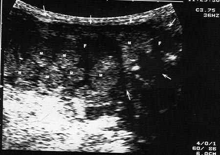
Figure 1: Ultrasound image of a dermoid cyst in the floor of the mouth. Ultrasound image obtained with a convex prob (3.75 Mhz), reveals the unilocular sac (arrows) full of globules of fat –the so called marbles (M)- floating within fluid matrix (F). |
 Büyütmek İçin Tıklayın |
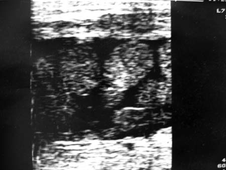
Figure 2: Ultrasound image of the same lesion obtained with linear prob (7.5 MHz) . |
Discussion
Most congenital dermoid cysts are thought to be arised due to an embryologic accident during the early stages of development, between 3 and 5 weeks of gestation [4]. The most popular theory regarding the etiology of this lesion suggests that it is derived from epithelial rests that become enclaved during midline closure of the first and second branchial arches.Dermoid cysts are frequently located in the midline, and are commonly found in the orbit, oral and nasal cavities and may occur as low as the suprasternal notch [1]. They are mobile and do not attach to the overlying skin. Unlike tyroglossal cysts, they do not have intimate relation with the hyoid bone and hence do not move with protrusion of the tongue. Clinically the cyst is palpated to be soft and well-encapsulated without associated lymphadenopathy. With puberty, its sudden increase in size was postulated to be related to the abundant sebum secretion from sebaceous glands.
Dermoid cysts are typically well-circumscribed, thin-walled, unilocular masses. Globules of fat floating within the lumen may produce a characteristic “sac-of-marbles” appearance on US. Globules of fat in the lumen appear as echogenic foci having acoustic shadowing. On the other hand, owing to various germinal layers, they may be heterogenous.
Dermoid cysts usually present in young adults. However, Bloom et al. [5] have reported the first case of a neonatal dermoid cyst in the floor of the mouth with extention to the midline of the neck. Although dermoid cysts are benign lesions, a case of malignant transformation to squamous cell carcinoma of a long-standing sublingual dermoid cyst is reported in the literature[6].
In differential diagnosis for midline cysts, thyroglossal duct cyst, inclusion cyst, cystic hygroma, ranula, nasal glioma and encephalocele (with cranial defects), blockage of the submandibular gland duct, neoplasms of the sublingual and minor salivary glands, neurofibroma, haemangioma and lymphangioma should be considered [1,7]. The main differential features of these lesions are their onset of age, location, and cystic contents.
In conclusion, a well-circumscribed, unilocular cystic lesion in the neck region, commonly in the floor of the mouth, appearing like a sac full of marbles on US examinations, highly suggest that the lesion is a dermoid cyst. This characteristic US feature should be taken into account in the differential diagnosis of benign neck masses.
Reference
1) Lev S, Lev MH. Imaging of cystic lesions. In: The radiologic clinics of North America. Philadelphia, Pennsylvania, W.B Saunders Company, 2000; 38:1013-1027. [ Özet ]
2) G. Ege, H. Akman, A. Senvar, G. Cakiroglu. Sublingual dermoid cyst, {Online} (2002.10.22). URL: http://www.eurorad.org/case.php?id=1787
3) Koeller KK, Alamo L, Adatr CF, Smirniotopoulos JG. Congenital cystic masses of the neck: Radiologic-pathologic correlation. Radiographics 1999;19:121-146. [ Özet ]
4) Smirniotopoulos JG, Chiechi MV. Teratomas, dermoids, and epidermoids of the head and neck. Radiographics 1995;15:1437. [ Özet ]
5) Bloom D, Carvalho D, Edmonds J, Magit A. Neonatal dermoid cyst of the floor the mouth extending to the midline neck. Arch Otolaryngol Head Neck Surg 2002;128:68-70. [ Özet ]
6) Devine JC, Jones DC. Carcinomatous transformation of a sublingual dermoid cyst. A case report. Int J Oral Maxillofac Surg 2000;29:126-127. [ Özet ]
7) Seah TH, Sufyan W, Singh B. Case report of a demoid cyst at the floor of the mouth. Ann Acad Med Singapore 2004;33(Suppl):77S-79S. [ Özet ]