


ENDONASAL ENDOSCOPIC RESECTION OF ETHMOID SINUS OSTEOMA WITH ORBITAL EXTENSION
Summary
Osteomas are the most common benign neoplasms of the nose and paranasal sinuses. They are commonly seen in the frontal sinus, rarely in the ethmoid sinuses. A 42-year-old female patient presented with intermittent frontal headache and blurred vision. Paranasal computed tomography scans revealed a bony mass in the left ethmoid sinus. Tumor was resected via the endoscopic endonasal approach. After surgical removal, patient became free of her symptoms. Follow-up of two years showed no recurrence.Introduction
Osteomas are the most common benign neoplasms of the nose and paranasal sinuses [1-6]. They are commonly asymptomatic, being an incidental finding in 1% of plain sinus radiographs [7,8] and 3% of computed tomography (CT) scans of the sinus [1,2,9,10]. They are commonly seen in the frontal sinus, less frequently in the ethmoid, rarely in the maxillary and quite exceptionally in the sphenoid sinuses [2,7,11,12,13]. They may occasionally grow rapidly affecting not only the paranasal sinuses, but also surrounding structures as well [7]. The symptoms include headache localized over the area of the osteoma, facial pain or deformity, rhinorrhea, anosmia, and sometimes sinusitis or ocular symptoms [1,9,14]. In this article we report a case of ethmoid osteoma presented with intermittent frontal headache and blurred vision and resected via endonasal endoscopic approach.Case Presentation
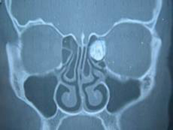
A 42-year-old woman presented with a one-year history of intermittent headache localized at the frontal area involving the glabella and dryness of nasal cavity especially in the left side. Two months before the presentation to our clinic, patient has presented ophthalmology department with the symptoms of left periorbital numbness and blurred vision of the left eye. She had a diagnosis of astigmatism and a pair of spectacles was recommended. When the ocular complaints remain the same and headache gets worse, she referred to the otolaryngology department. In physical examination there was no facial deformity, globe protrusion or limited eye movements. The other clinical findings were normal. Coronal paranasal sinus CT revealed a 2x1 cm bony mass in the left anterior ethmoid cells (Fig.1).
 Büyütmek İçin Tıklayın |
Figure 1: Coronal paranasal sinus CT revealed a 2x1 cm bony mass in the left anterior ethmoid cells |
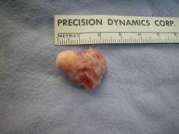
The mass which was considered as osteoma was removed by dissecting from surrounding structures with endonasal endoscopic approach (Fig.2). A bony defect was determined in lamina papyricea. Histopathological examination confirmed the diagnosis of osteoma.
 Büyütmek İçin Tıklayın |
Figure 2: Osteoma removed via endonasal endoscopic approach. |
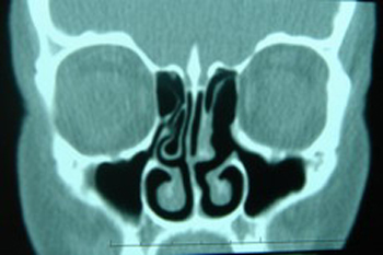
The postoperative course of the patient was uneventful. The headache complaint of the patient improved immediately after the surgery. The ocular symptoms of the patient resolved completely in seven days, so that she had no need to use spectacles anymore. Coronal paranasal sinus CT six months after the surgery demonstrated a minimally defective lamina papyricea in the left side and no sign associated with recurrence (Fig.3). At present, the patient is free from her symptoms for two years.
 Büyütmek İçin Tıklayın |
Figure 3: Postoperative coronal paranasal sinus CT demonstrated no sign associated with recurrence. |
Discussion
Osteomas of the paranasal sinuses are generally small and characteristically asymptomatic [2,7,9,11,13,15,16]. The most common symptom of a paranasal sinus osteoma is headache localized over the area of the osteoma [9,15,17-19]. The most common ocular findings are proptosis, extraoculer muscle displacement, optic disk edema, choroidal folds, and orbital infection [5]. Other ocular findings include shooting retrobulbar pain and transient visual disturbances. In this case patient presented primarily with headache. Patient was suffering also ocular symptoms like periorbital numbness and blurred vision. Although the physical examination of the patient did not reveal a clinically evident proptosis, a defect in lamina papyricea following the removal of the osteoma which is an objective sign for the orbital extension may explain the blurred vision.Plain-film radiography is the basis of the diagnosis of osteoma [20]. Osteomas are best visualized with CT [21]. Computed tomography is very important in the evaluation of bone extension of the tumor; also the site of origin (paranasal sinuses, orbital roof, ethmoid and sphenoid bone) may be often defined on the coronal scans [12]. In this case the diagnosis of ethmoid sinus osteoma achieved by CT findings.
The management of ethmoid osteomas remains controversial [1,9]. Generally osteomas treated conservatively. No treatment is recommended for asymptomatic osteomas, especially in elderly subjects [2,9,12,22]. Indications for surgery in paranasal osteomas include sphenoid osteomas, irrespective of size, presence of significant symptoms like unexplained headache, recurrent sinusitis, ocular symptoms, central nervous system symptoms, enlargement seen radiographically, extension beyond confines of the sinus, filling of more than 50% of the volume of the frontal sinus, location near the frontal sinus ostium, cosmetic deformity [7,9]. In this report patient presented with unexplained intermittent headache and ocular symptoms. Therefore, she was a good candidate for surgical treatment.
The surgical approach must protect the vital structures, while optimizing the ability to totally excise the osteoma with minimal cosmetic deformity [7,9,11]. For large osteomas of the ethmofrontal region, surgical excision, including the osteoplastic flap technique, lateral rhinotomy, or direct anterior surgical exposure were used whenever there was evidence of progressive growth and involvement of surrounding structures [1]. A dacryocystorhinostomy approach using a mastoid drill and perforating burr has been described [1,23]. Transcoronal removal with an osteotome has been also described [1,11]. The necessities of these open procedures for slow growing, benign, and encapsulated tumors continue to be debated [1]. In well-selected cases, endoscopic sinus surgery offers a convenient, safe, and effective alternative to open procedures with reduced morbidity and superior cosmetic results [1,2,9,24,25]. In this case the endonasal endoscopic approach was used and ethmoid sinus osteoma dissected from surrounding structures. Patient was discharged immediately after the operation.
Conclusion
Etmoid sinus osteomas can cause headache and ocular symptoms. In suspicious cases radiological examination especially paranasal sinus CT will help the diagnosis. In symptomatic cases osteoma should be resected. In well-selected cases, endoscopic sinus surgery offers a convenient, safe, and effective alternative to open procedures with reduced morbidity and superior cosmetic results.Reference
1) Huang HM, Liu CM, Lin KN, Chen HT. Giant ethmoid osteoma with orbital extension, a nasoendoscopic approach using an intranasal drill. Laryngoscope 2001; 111(3):430-2. [ Özet ]
2) Naraghi M, Kashfi A. Endonasal endoscopic resection of ethmoido-orbital osteoma compressing the optic nerve. Am J Otolaryngol 2003;24(6):408-412. [ Özet ]
3) Hehar SS, Jones NS. Fronto-ethmoid osteoma: the place of surgery. J Laryngol Otol 1997; 111(4):372-375. [ Özet ]
4) Aldren CP. Bony remodelling in an osteoma of the paranasal sinuses. J Laryngol Otol 1993;107:633-635. [ Özet ]
5) Schwartz MS, Dennis M. Crockett. Management of a large frontoethmoid osteoma with sinus cranialization and cranial bone graft reconstruction. Int J Pediatr Otorhinolaryngol 1990;20(1):63-72. [ Özet ]
6) Gökçeer T, Kahve, Naiboglu B, Atbaş A. Ethmoid sinus osteoma with orbital extension. Kulak Burun Boğaz İhtis Derg 2003;10(3):117-120. [ Özet ]
7) Savic DL, Djeric DR. Indications for the surgical treatment of osteomas of the frontal and ethmoid sinuses. Clin Otolaryngol 1990; 15(5): 397-404. [ Özet ]
8) Harrison D. (1979) Tumors of the nose and sinuses. In Scott-Brown’s Disease of the Ear, Nose, Throat, The Nose and Sinuses, pp.367-369. Butterworths, London.
9) Mansour AM, Salti H, Uwaydat S, Dakroub R, Bashshour Z. Ethmoid sinus osteoma presenting as epiphora and orbital cellulitis: Case report and literature review. Surv Ophthalmol 1999;43(5):413-426. [ Özet ]
10) Earweaker J. Paranasal sinus osteomas: a review of 46 cases. Skeletal Radiol 1993;22:417-423. [ Özet ]
11) Marks M, Newman H. Transcoronal removal of an atypical orbitoethmoid osteoma. Plast Reconstr Surg 1983;72(6):874-7. [ Özet ]
12) Maiuri F, Iaconetta G, Giamundo A, Stella L, Lamaida E. Fronto-ethmoidal and orbital osteomas with intracranial extension: report of two cases. J Neurosurg Sci 1996;40(1):65-70.
13) Koyuncu M, Belet Ü. Huge osteoma of the frontoethmoidal sinus with secondary brain abscess. Auris Nasus Larynx 2000;27:285-287. [ Özet ]
14) Van Manen SR, Bosch DA, Peeters FL, Troost D. Giant intracranial mucocele. Clin Neurol Neurosurg 1995;97:156-160.
15) Koivunen P, Löppönen H. The growth rate of osteomas of the paranasal sinuses. Clin Otolaryngol 1997;22:111-114. [ Özet ]
16) Manaka H, Tokoro K. Intradural extension of mucocele complicating frontoethmoid sinus osteoma: case report. Surg Neurol 1998;50:453-456. [ Özet ]
17) Smith ME, Calcaterra TC. Frontal sinus osteoma. Ann Otol Rhinol Laryngol 1989;98:896-900. [ Özet ]
18) Boysen M. Osteomas of the paranasal sinuses. J Otolaryngol 1978;7:366-370. [ Özet ]
19) Mugliston TA, Stafford N. A cranio-facial approach to large osteomas of the fronto-ethmoidal region. J Laryngol Otol 1985;99(10):979-983. [ Özet ]
20) Greenspan A. Benign bone-forming lesions: osteoma, osteoid osteoma and osteoblastoma. Clinical, imaging, pathologic, and differential considerations. Skeletal Radiol 1993;22(7):485-500. [ Özet ]
21) Grayeli AB, Redondo A, Sterkers O. Anterior skull base osteoid osteoma: case report. Br J Neurosurg 1998; 12(2):173-5. [ Özet ]
22) Al-Sebeih K, Desrosiers M. Bifrontal endoscopic resection of frontal sinus osteoma. Laryngoscope 1998;108:295-298. [ Özet ]
23) Soboroff BJ, Nykiel F. Surgical treatment of large osteomas of the ethmofrontal region. Laryngoscope 1996;76:1068-1081. [ Özet ]
24) Cronin ED, Ruiz-Razura A, Livingstone CK, et al. Endoscopic approach for the resection of forehead masses. Plast Reconstr Surg 2000;105:2459-2463. [ Özet ]
25) Menezes CA, Davidson TM. Endoscopic resection of a spheno-ethmoid osteoma: a case report. Ear Nose Throat J 1994;73:598-600. [ Özet ]